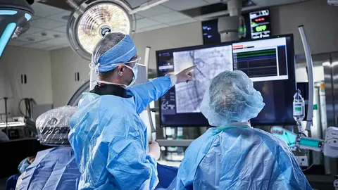
Interventional radiology uses different imaging types to diagnose and treat health conditions. Imaging helps precisely locate blood vessels, organs, and soft tissues, supporting various kinds of treatment plans. During treatment, instead of large incisions, specialists use imaging to reach a specific area through a small entry point in the skin. Here’s more information on the benefits of imaging and how interventional radiology specialists use it to guide your care:
Providing Musculoskeletal Imaging
Musculoskeletal imaging focuses on bones, joints, muscles, tendons, and ligaments, using several technologies to guide healthcare decisions. Each modality has different strengths. X-rays show bone alignment and joint spaces, while CT scans provide more detailed cross-sectional views of complex structures. MRI scans use magnetic fields and radio waves to highlight soft tissues, often aiding diagnosis. Ultrasound uses sound waves to visualize moving structures, such as tendons or blood vessels, in real time.
Imaging helps identify fractures, ligament tears, tendon injuries, and degenerative joint changes, and it also helps track how these issues change over time. When interventional radiology treatments are needed, imaging guides needles or other tools to specific body areas. MRI and ultrasound highlight inflammation or fluid collections in soft tissues, while CT and X-ray outline structural changes affecting movement. Because imaging shows the exact path of a needle or catheter, procedures stay focused on the problem area while sparing nearby structures.
Guiding Angiography Procedures
Angiography uses contrast dye and X-ray imaging to study blood vessels in detail to support condition diagnosis and treatment planning. A provider may recommend this test when there are questions about blood flow, vessel narrowing, or structural changes inside arteries and veins. During this procedure, a catheter is threaded into the vascular system, and imaging tracks its position as contrast flows through the arteries or veins. This lets specialists see where blood flows freely and where it slows or stops. These images support decisions about next steps, such as medication management, surveillance, or an image-guided intervention.
Supporting Other Procedures
Imaging guidance supports a range of interventional radiology procedures beyond angiography, helping keep many treatments focused and efficient. During drainage procedures, ultrasound or CT helps locate fluid collections and guide a catheter into the safest path, reducing the need for larger incisions. Biopsies often rely on live imaging to guide a needle to a specific lesion, supporting tissue sampling from hard-to-reach areas. Tumor ablation and cryoablation also rely on CT, ultrasound, or MRI to confirm probe placement, monitor the treatment zone, and protect nearby organs or nerves. Because imaging is used throughout these procedures, care teams can adjust treatment in real time as needed. This level of visualization supports personalized care plans built around each patient’s anatomy.
Learn More About Interventional Radiology
Imaging-guided care gives patients and providers a shared view of what is happening inside the body. Using real-time visualization, interventional radiology combines diagnostic imaging and treatment into a single, coordinated approach. To learn how these procedures relate to your healthcare needs, contact a radiology clinic near you for personalized medical advice. Learning more about these options helps you stay informed about your diagnostic and treatment plans.
